With modern lifestyles often involving long hours of sitting and unbalanced diets, the risk of cardiovascular diseases is steadily increasing. Have you ever experienced numbness, swelling, or pain in your limbs? Or suspected a possible vascular blockage? In these situations, vascular ultrasound is a fast, painless, and effective diagnostic tool.
This technique uses sound waves to evaluate blood flow in veins and arteries throughout the body. By observing how blood moves through the vessels, doctors can assess for possible issues such as deep vein thrombosis (DVT), arterial narrowing, blockages, or other vascular abnormalities. Combined with mated measurement tools, vascular ultrasound enables rapid, accurate assessments—especially valuable in emergency rooms, outpatient clinics, and pre- or post-operative evaluations.
What Can Vascular Ultrasound Examine?
Blood vessels are found all over the body. Using vascular ultrasound, clinicians can assess:
- Carotid arteries
- Veins and arteries in upper and lower limbs
- Abdominal or peripheral arteries
Key conditions it can detect include:
- Thrombosis (e.g., DVT – deep vein thrombosis)
- Vascular stenosis or occlusion (e.g., carotid artery narrowing)
- Abnormal blood flow velocities (analyzed via PSV, EDV, RI, and PI indicators)
- Venous insufficiency (e.g., varicose veins)
- Pseudoaneurysms or vessel ruptures
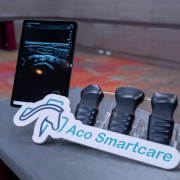
Why Choose a Handheld Ultrasound?
While traditional ultrasound systems offer high-resolution imaging and full-feature capabilities, handheld ultrasound devices provide unmatched portability, faster scanning, and app integration—ideal for tight spaces or outdoor environments. These features make handheld systems a powerful tool for point-of-care vascular assessment.
How Does Aco Smartcare’s Apache App Help?
In vascular evaluations, capturing Doppler waveforms and calculating indicators has traditionally been a time-consuming process. The Apache App by Aco Smartcare simplifies this with smart features like:
- Auto Trace
- Stenosis%
- PI/RI
These tools allow handheld ultrasound devices to go beyond convenience—offering real-time assistance for accurate interpretation, making them an essential asset in modern clinical workflows.
(Related Reading:Aco Smartcare – Vascular Flow Analysis)
Are There Risks with Vascular Ultrasound?
Unlike invasive angiography or costly imaging modalities, vascular ultrasound is non-invasive, radiation-free, and safe for repeated use. This makes it ideal for long-term monitoring, especially in chronic conditions like diabetes or kidney disease. It’s a powerful tool for early detection and prevention of cardiovascular complications.
For healthcare professionals, it provides real-time visualization of blood flow, direction, velocity, and waveform changes—enabling faster diagnosis of blockages, stenosis, thrombosis, or perfusion abnormalities. Even in remote or resource-limited settings, it empowers clinicians to act quickly without needing to wait for bulky equipment.
(Related Reading:Apache neo L154 Handheld Ultrasound)
References data
<RadiologyInfo.org – Ultrasound – Vascular>
<Vascular Ultrasound – AtriumHealth>